Calcium
dynamics in the 3D space of the cardiac myocyte
Pavol Petrovič*1, Alexandra Zahradníková2,
Ivan Valent1
1Faculty of Natural Sciences, Comenius University, Bratislava, Slovakia
2Institute of Molecular
Physiology and Genetics, Slovak Academy of Sciences, Bratislava, Slovakia
The dynamics
of cytosolic calcium plays an essential role in cardiac myocyte contraction and
it also has an essential impact on electrical activity of the myocyte. The spontaneous calcium
release from sarcoplasmic reticulum via RyR channels during the diastole causes
calcium sparks that may lead to formation and propagation of calcium waves
under certain conditions. The mechanism of the propagation of those waves is
probably determined by the interaction between calcium-induced calcium release (CICR), calcium diffusion, and calcium reuptake.
In the presented
work we describe several models in 2D and 3D space, analyzing calcium dynamics
in the cytosol. Calcium release description was based on gating of RyR channels
using the aHTG gating scheme (Zahradníková,
this meeting). Calcium diffusion and reuptake was described using a
stochastic generalization of the fire-diffuse-fire (FDF) framework (Coombes at al., 2004). The diffusion from
distributed sources can, in principle, be computed by convolving these
point-source solutions over a known source distribution. In our models, we used
the fast and convenient method for treating distributed sources by
reformulating the problem with Fast Fourier Transforms algorithm (Coombes
at al., 2004).
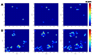
Activation
of calcium sparks (Fig. 1A) was found to be dependent on the number of
activated channels in the RyR clusters (CRU- calcium release unit) as well as
on the cytosolic Mg2+ concentration, yet not sensitive to the
arrangement of the CRU on the surface. We can observe saltatory
or continuous waves (Fig. 1B) propagation in the 2D model, depending on the
model parameters and on the CRU arrangement. In the 3D model, wave generation was
difficult to be attained under the same conditions as in 2D. After and increase
of the diffusion coefficient and the calcium flux from CRU we can detect the
waves as well.
|
|
Fig. 1.
Snapshots in 2D generated every 15 ms for (A) spark formation (D = 30µm2/s, JCa = 2.5µM µm) and (B) wave generation and
propagation (D = 30µm2/s, JCa = 15.5µM µm). |
References
Coombes, S, Hinch, R., Timofeeva, Y. Receptors, sparks and waves in a fire-diffuse-fire framework for calcium release. Progress in Biophysics & Molecular Biology 85: 197-216. 2004